Biology · Class 11 / NEET & CBSE
Locomotion & Movement: Sliding Filament Theory of Muscle Contraction
Master muscular and skeletal physiology: Sarcomere microanatomy (A-band, I-band, H-zone, Z-disc), Actin (Troponin/Tropomyosin) and Myosin filaments, Neuromuscular junction, and Calcium-ATP cross-bridge cycle.
11. Visual Overview & Intuitive Discovery: Locomotion & Movement: Sliding Filament Theory of Muscle Contraction
🔍 Clickable Interactive Study Prompts (Click to explore or jump to section):
Clickable### Intuitive Foundations & Real-World Reality
In 1954, Hugh Huxley and Jean Hanson published simultaneous papers in Nature revealing the Sliding Filament Theory. Muscles do not contract by shrinking or coiling protein molecules like springs; rather, thick filaments (Myosin) and thin filaments (Actin) slide past one another through repeating cycles of ATP-powered cross-bridge rowing, shortening the functional contractile unit—the Sarcomere.
 Full size
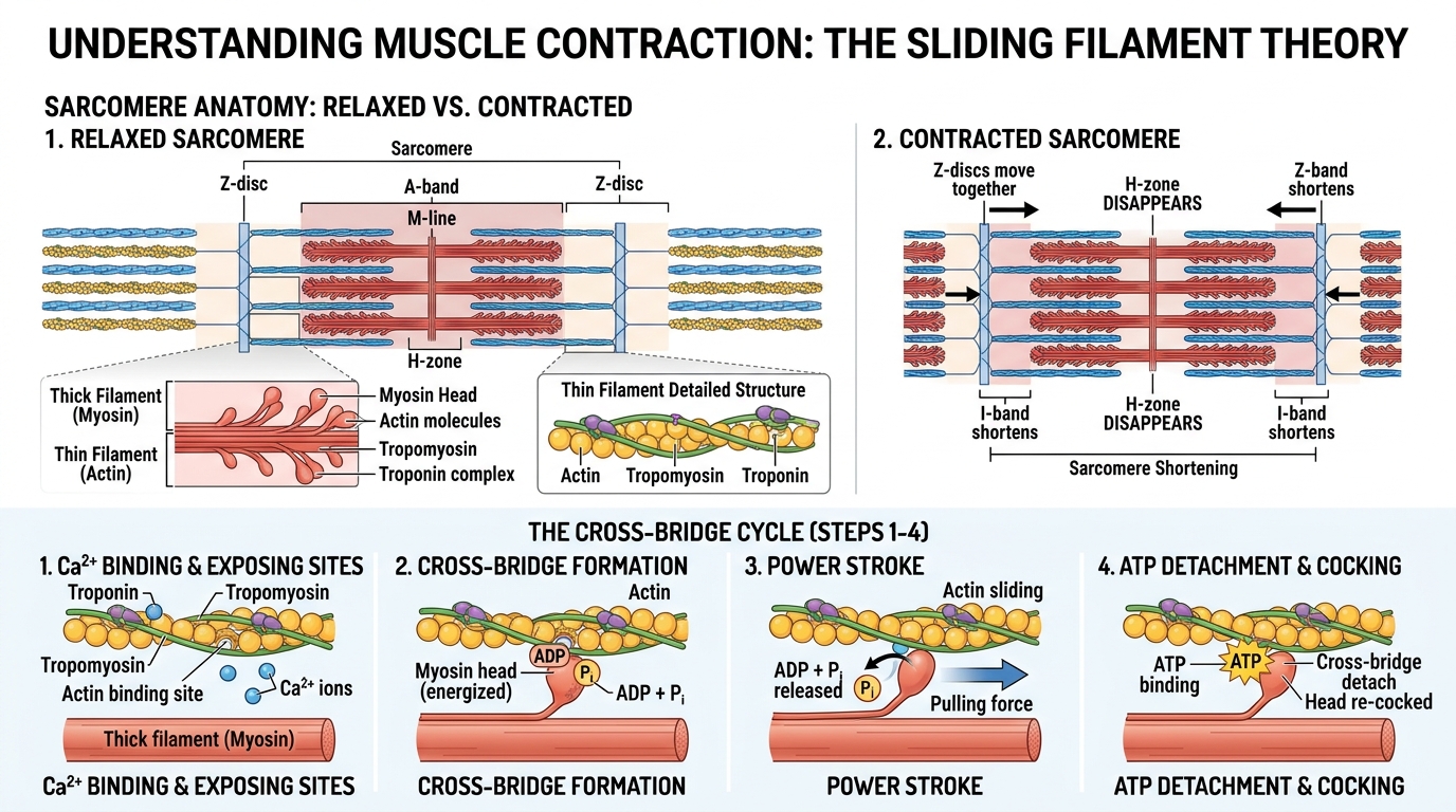
Sliding Filament Theory & Sarcomere Contraction
Detailed sarcomere anatomy (A-band, I-band, H-zone, Z-discs), relaxed vs contracted state, and the 4-step cross-bridge cycle with ATP and Ca2+.
- 1Core Anchor 1 — Sarcomere Structure: A-Band, I-Band, H-Zone, Z-Line & M-Line
- 2Core Anchor 2 — The Calcium Switch: Ca²⁺ Binds Troponin C to Unmask Myosin Binding Sites
- 3Core Anchor 3 — Cross-Bridge Cycle: ATP Hydrolysis, Power Stroke & ATP Detachment
- 4Core Anchor 4 — Rigor Mortis: Biochemical Cause of Post-Mortem Muscle Stiffness
22. Concept & Decision Flowchart: Neuromuscular Excitation & Cross-Bridge Flowchart
Neuromuscular Excitation & Cross-Bridge Flowchart
- 1
1. Neuromuscular Action Potential
Motor neuron fires action potential -> Acetylcholine released at motor endplate -> triggers muscle action potential down T-tubules.
- 2
2. Calcium Release from Sarcoplasmic Reticulum
Depolarization opens Ryanodine receptor Ca²⁺ release channels -> cytosolic Ca²⁺ levels soar from 10⁻⁷ M to 10⁻⁵ M.
- 3
3. Unmasking Active Sites
Ca²⁺ binds Troponin C -> conformational change pulls Tropomyosin away from active binding sites on Actin filaments.
- 4
4. Cross-Bridge Cycling & Power Stroke
Myosin head (energized by ATP hydrolysis to ADP + Pi) binds actin -> releases Pi to execute Power Stroke (pulls actin toward M-line) -> fresh ATP binds to detach myosin head. Cycle repeats!
33. Step-by-Step Structural Dynamics: Dimensional Changes in Sarcomere Bands During Contraction
Derivation — Step-by-Step Structural Dynamics: Dimensional Changes in Sarcomere Bands During Contraction
- 1
Actin thin filaments anchor into the elastic Z-disc.
- 2
Length of myosin thick filaments remains unchanged.
- 3
Thin filaments overlap deeply between thick filaments.
- 4
Opposing actin filaments meet and overlap at the central M-line.
44. Real-World Case Study & Industrial Application
### Forensic Pathology: Rigor Mortis and Time of Death Estimation
Following clinical death, cellular respiration terminates and ATP production plunges to zero. In the absence of ATP, the Sarcoplasmic Reticulum cannot pump Ca²⁺ back, leaving troponin unmasked. Simultaneously, Myosin heads cannot detach from Actin filaments because detachment strictly requires fresh ATP binding. Myosin heads remain locked in a perpetual cross-bridge state (Rigor Mortis), stiffening the corpse for 12-24 hours until lysosomal enzymes autodigest muscle proteins.
55. High-Yield Reference Data & Solved Problems
High-Yield Comparison & Key Constants
| Sarcomere Region | Filament Composition | Behavior During Muscle Contraction |
|---|---|---|
| Sarcomere Length (Z to Z) | Complete contractile unit | Shortens significantly |
| A-Band (Dark Band) | Thick myosin + overlapping actin | Remains completely UNCHANGED in length |
| I-Band (Light Band) | Thin actin filaments strictly | Shortens (pulled inward toward center) |
| H-Zone | Central region of thick myosin only | Shortens and completely DISAPPEARS |
| Z-Discs | Protein anchor boundary plate | Pulled closer together toward M-line |
Sarcomere Length (Z to Z)
- Filament Composition
- Complete contractile unit
- Behavior During Muscle Contraction
- Shortens significantly
A-Band (Dark Band)
- Filament Composition
- Thick myosin + overlapping actin
- Behavior During Muscle Contraction
- Remains completely UNCHANGED in length
I-Band (Light Band)
- Filament Composition
- Thin actin filaments strictly
- Behavior During Muscle Contraction
- Shortens (pulled inward toward center)
H-Zone
- Filament Composition
- Central region of thick myosin only
- Behavior During Muscle Contraction
- Shortens and completely DISAPPEARS
Z-Discs
- Filament Composition
- Protein anchor boundary plate
- Behavior During Muscle Contraction
- Pulled closer together toward M-line
Exam question · JEE Main, Advanced & NEET Standard
- 1. ATP Hydrolysis: Myosin head possesses ATPase activity. It hydrolyzes ATP into ADP and Inorganic Phosphate (Pi), entering a high-energy cocked conformation.
- 2. Cross-Bridge Formation: When Ca²⁺ binds Troponin C, tropomyosin shifts to expose actin binding sites. The energized myosin head binds to the exposed actin active site.
- 3. The Power Stroke: Release of Inorganic Phosphate (Pi) triggers the conformational tilting of the myosin head (by 45°), pulling the actin filament toward the center of the sarcomere (M-line). ADP is released during this stroke.
- 4. Cross-Bridge Detachment: A new molecule of ATP binds to the myosin head, causing the myosin-actin cross-bridge to detach.
- 5. Reactivation: The fresh ATP is hydrolyzed to ADP + Pi, re-cocking the head for the next contractile cycle.
Answer: 1. ATP Hydrolysis: Myosin head possesses ATPase activity. It hydrolyzes ATP into ADP and Inorganic Phosphate (Pi), entering a high-energy cocked conformation.
2. Cross-Bridge Formation: When Ca²⁺ binds Troponin C, tropomyosin shifts to expose actin binding sites. The energized myosin head binds to the exposed actin active site.
3. The Power Stroke: Release of Inorganic Phosphate (Pi) triggers the conformational tilting of the myosin head (by 45°), pulling the actin filament toward the center of the sarcomere (M-line). ADP is released during this stroke.
4. Cross-Bridge Detachment: A new molecule of ATP binds to the myosin head, causing the myosin-actin cross-bridge to detach.
5. Reactivation: The fresh ATP is hydrolyzed to ADP + Pi, re-cocking the head for the next contractile cycle.
What it tests: Tests rigorous conceptual mastery, dimensional analysis, and multi-step deduction.
Remember
- Locomotion & Movement: Sliding Filament Theory of Muscle Contraction: Foundational law governing physical, chemical, and biological phenomena.
- Mastered derivation: Step-by-Step Structural Dynamics: Dimensional Changes in Sarcomere Bands During Contraction.
- Avoided classic trap: During muscle contraction, protein filaments DO NOT shorten! Neither actin nor myosin shrinks in length. They ...
- Real-world relevance: Proven clinical, aerospace, and industrial significance.
- High-yield exam rule: Always verify dimensional consistency and physical boundary conditions.
Check yourself
Question 1
What is the primary governing principle of Locomotion & Movement: Sliding Filament Theory of Muscle Contraction?Question 2
In competitive examinations (JEE/NEET), what is the most critical pitfall in Locomotion & Movement: Sliding Filament Theory of Muscle Contraction?